For most of us, going to the doctor can be unpleasant, but, over the years, medicine has evolved so much and at such a rapid pace that almost any kind of disease, spotted at the right time, can be treated and even cured.
MDs have a hard time explaining to patients that the right examination can save their life because the fear of the unknown and the fear of medical procedures can be overwhelming for the patient. One of the most common worries most of us have is undergoing an endoscopy.
But if you know more about its history and about the endoscope, as well as how it evolved over time, you will be happy that this medical device exists, and that it can be used for your examination. Moreover, it can even save your life.
What are endoscopes used for?
An endoscopy is a procedure that enables MDs to look inside the human body. You might think that a medical device that can do that was invented only in the twentieth century, but you will be surprised to find out that a similar instrument has been found in Pompeii’s ruins, meaning that the ancient Romans used some kind of similar device as well.
A short history
In 1805, Phillip Bozzini realized that for treating his patients he needed a device to look inside their urinary tract, pharynx, and rectum. So, he invented the Lichteiter, a tube that was used to light up the examined area.
After that, in 1853, to examine the bladder and the urinary tract, a french MD, by the name of Antoine Jean Desormeaux, invented an instrument called an endoscope. This was the first time in history that the device got its name which is still used today.
After various MDs used it to examine the bladder, they started wondering if, perhaps, it would be possible to use it for other organs as well. Therefore, in 1868, a German doctor, Adolph Kussmaul, managed to look inside a patient’s stomach.
After more than a decade, in 1881, Dr. Johnatan von Mikulicz developed an instrument that is now known as a rigid gastroscope, the first one that had the purpose to be used for practical applications.
However, MDs using the device realized that they need a more practical and ergonomic one. One of the most significant problems was that the endoscope was not flexible and the pain that the patient would be exposed to was excruciating.
But, finally, in 1932, a doctor named Rudolph Shindler modified the original version and developed a gastroscope that was flexible. He added some lenses so that he could have a maximized view of the patients’ stomachs.
Over time, technology evolved and MD’s started to envision the possibility to take pictures inside the body and examine them even further, keeping them as history into the patients’ medical records.
So, in 1949, a doctor working for the medical center at Tokyo University made a proposal to an optical company to invent a camera that would be small enough to be able to reach the stomach attached to an endoscope. It was a tough job for researchers because a photography camera is a complex device that includes a lot of lenses.
The production of such small lenses was a challenge for researchers, but also the illumination part. They eventually figured it out, and developed a camera with the most important three features: good image quality, reduced discomfort for the patient, and it could also photograph all the sides of the stomach.
What about now?
As time passed, researchers all over the world tried to develop a better endoscope. Over the years, they started using glass fiber, a material that enabled the light to pass all over the tube even when it was bent.
Being able to make real-time observations was a big breakthrough for doctors, but this device couldn’t take photos. It wasn’t until the 60s, when the live camera was invented and it made a significant difference for doctors and patients all over the world.
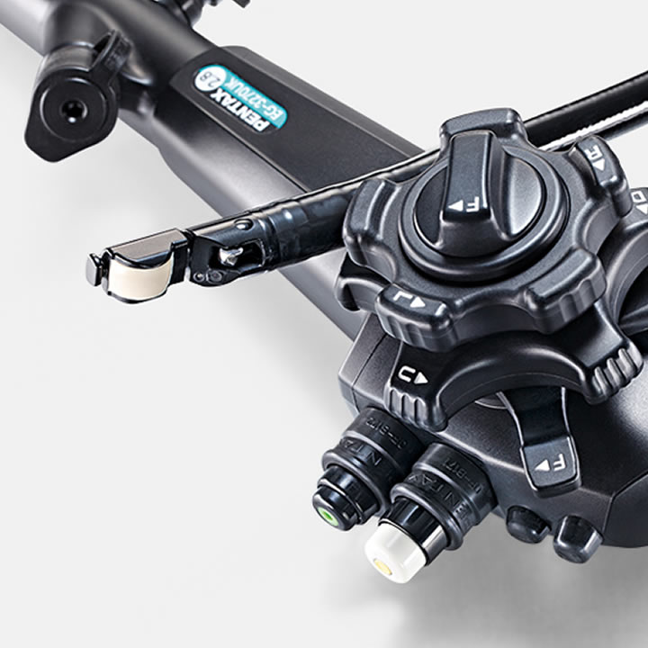
Nowadays, MDs use endoscopes for live examinations of patients’ organs, to film them, store them as medical history and even perform procedures like ulceration cauterization, where they have to use a waterproof one.
They can detect tumors at an early stage and detect formations in the stomach, colon, and rectum. In just a few minutes, an endoscope can deliver images from inside the body that can help doctors diagnose and treat patients at a higher rate and save millions of lives.
We hope this information changed the way you look at the endoscopy procedure and realize that, over time, researchers developed instruments that produce minimal discomfort and that you are very lucky to be living in the 21st century. The endoscope is an amazing invention that should be appreciated at its true value.

